Page Not Found
Page not found. Your pixels are in another canvas.
A list of all the posts and pages found on the site. For you robots out there is an XML version available for digesting as well.
Page not found. Your pixels are in another canvas.
This is a page not in th emain menu
Published:
In 2020, we started a spin-out company Infinite, Biomedical Technologies, devoted to animal-free safety testing of nanomaterials and chemicals. We are excited to pursue this joint journey to bridge all TRL levels from basic science to the final product/service.
Published:
LBF has a long tradition in development of technologies and infrastructure in the fields of tissue engineering, antimicrobial surfaces, biomedical and pharmaceutical applications, optimization of industrial materials and processes, and reasonable use of energy and building biophysics.
Published:
The editors of Advanced Materials selected our recent paper on the prediction of chronic inflammation for the frontispiece of the issue!
Published:
Our spin-out company Infinite Biotech d.o.o. is going live! We will revolutionise safety testing for nanomaterials and chemicals by new in vitro approaches. Stay tuned!
Published:
Our MSc student Urša Uršič brilliantly defended her thesis! Congratulations and all the best with her PhD at the Max Planck Institute of Molecular Cell Biology and Genetics in Dresden, Germany!
Published:
Our paper on reliable labelling of nanoparticles has just been published in Nanotoxicology! Congrats, Boštjan & Hana!
Published:
We are searching for talented and motivated people to join our team! Currently we are seeking staff with a physics-related backround for development of microscope-on-chip technologies.
Published:
Our MSc student Ana Jandrlić defended her thesis today at the Faculty of Pharmacy, University of Ljubljana - congratulations, and welcome to the team of our spin-out company Infinite!
Published:
Our MSc student Petra Čotar defended her thesis at the Faculty of mathematics and physics today! Congratulations, we’re glad you stay in our team for further adventures!
Published:
Our MSc student Tanja Vajs defended her thesis today at the Faculty of natural science and mathematics, University of Maribor. Congratulations, Tanja - we wish you all the best with your future undertakings!
Published:
Four enthusiastic students from the MSc programme Laboratory Biomedicine (Faculty of Pharmacy, University of Ljubljana) join our team. Looking forward to overcoming exciting scientific challenges with fresh forces!
Published:
Would you like to co-create diagnostic technologies of the future? Join our team!
Published:
Hana Kokot defended her PhD thesis today at the Jozef Stefan International Postgraduate School - woohoo!!
Published:
Darin Lah is starting his PhD journey between our lab and the spin-out company Infinite. Looking forward to exciting new research with fresh ideas!
Published:
Today we launched the EU project nanoPASS that we coordinate!
Published:
Our team will be expanding with a diligent part-time lab technician!
Published:
We are searching for a talented and motivated postdoc to join our team and work on the newly started EU project nanoPASS! The project is devoted to the development of new methods for safety testing of nanomaterials, which will be faster, cheaper and more reliable than in vivo tests. Our greatest challenge is to predict slowly developing diseases, such as lung fibrosis, cardiovascular disease, and neurodegeneration.
Published:
Boštjan Kokot defended his PhD thesis today at the University of Maribor!
Published:
Within nanoPASS we organised a consultation with local experts in toxicology, environmental science, health practitioners, and regulators. It has been very instructive to learn the views from all sides.
Published:
Our team will be expanding with a diligent expert associate to work in our biological and chemical lab.
Published:
Cody is joining the team to build microscopes, analyse images, mess with nanoparticles, and more. Looking forward to solving big nanoscale problems together!
Published:
Benjamin Koroševič Koser is starting his PhD project with us, focusing on the prediciton of cancer induced by inhalation of particulate matter.
Published:
Aleksandar Sebastijanović defended his PhD thesis “In Vitro-Detected Early Events to Unravel Sporadic Neurodegeneration” today at the Jozef Stefan International Postgraduate School! It was a joy to witness such a rich discussion - going both deep into mechanisms as well as broad into implications.
Published:
Our MSc students Ana Vencelj, Tamara Picaboo Kuster, and Matic Zupančič defended their theses at the Faculty of pharmacy, University of Ljubljana. Congratulations and thanks for having contributed to our efforts to understand triggering of long-term diseases by particulate matter!
Published in European Biophysics Journal, 2009
As proteins are key molecules in living cells, knowledge about their structure can provide important insights and applications in science, biotechnology, and medicine. However, many protein structures are still a big challenge for existing high-resolution structure-determination methods, as can be seen in the number of protein structures published in the Protein Data Bank. This is especially the case for less-ordered, more hydrophobic and more flexible protein systems. The lack of efficient methods for structure determination calls for urgent development of a new class of biophysical techniques. This work attempts to address this problem with a novel combination of site-directed spin labelling electron spin resonance spectroscopy (SDSL-ESR) and protein structure modelling, which is coupled by restriction of the conformational spaces of the amino acid side chains. Comparison of the application to four different protein systems enables us to generalize the new method and to establish a general procedure for determination of protein structure.
Download here
Published in Biophysical journal, 2010
To characterize the structure of dynamic protein systems, such as partly disordered protein complexes, we propose a novel approach that relies on a combination of site-directed spin-labeled electron paramagnetic resonance spectroscopy and modeling of local rotation conformational spaces. We applied this approach to the intrinsically disordered C-terminal domain of the measles virus nucleoprotein and provided a mechanistic explanation for the high affinity of the binding reaction between the measles virus and the viral phosphoprotein.
Download here
Published in Cell Biology International, 2010
We characterized the influence of three different detachment procedures: cell scraping by rubber policeman, trypsinization and a citrate buffer treatment on V-79 cells in the plateau phase of growth (arrested in G1). We found out that cell viability was 91% for trypsin treatment, 85% for citrate treatment and 70% for cell scraping.
Download here
Published in Organic & Biomolecular Chemistry, 2011
A serious drawback of ESR, particularly in its application to cells, is the lack of information on the location of spin probes in the system. In order to realize real time tracking, a spin probe was combined with a fluorophore in a new kind of nitroxide–fluorophore double probe which, in addition to information about lipid dynamics, enables visualization by fluorescence microscopy.
Download here
Published in Biomedical optics express, 2011
Here we present a development of fluorescence microspectroscopy (spectral imaging) based on a white light spinning disk confocal microscope with emission wavelength selection by a liquid crystal tunable filter. Spectral contrasting of images was used to localize polymer nanoparticles and cell membranes labeled with fluorophores that have substantially overlapping spectra. In addition, fluorescence microspectroscopy enabled spatially-resolved detection of small but significant effects of local molecular environment on the properties of environment-sensitive fluorescent probe.
Download here
Published in Biophysical journal, 2013
In this study, bleaching-corrected polarized fluorescence microspectroscopy with nanometer spectral peak position resolution was applied to characterize conformations of two alkyl chain-labeled 7-nitro-2-1,3-benzoxadiazol-4-yl phospholipids in three model membranes, representing the three main lipid phases. The combination of polarized and spectral detection revealed two main probe conformations with their preferential fluorophore dipole orientations roughly parallel and perpendicular to membrane normal. Their peak positions were separated by 2–6 nm because of different local polarities and depended on lipid environment.
Download here
Published in Optics express, 2013
Fluorescence microspectroscopy (FMS) with environmentally sensitive dyes provides information about local molecular surroundings at microscopic spatial resolution. We showed that careful analysis of data acquired by stochastic wavelength sampling enables nanometer spectral peak position resolution even for highly photosensitive fluorophores with small spectral shifts, which are commonly used in bulk spectrofluorimetry but largely avoided in microspectroscopy.
Download here
Published in J. Chem. Phys., 2014
All atom molecular dynamics (MD) models provide valuable insight into the dynamics of biophysical systems, but are limited in size or length by the high computational demands. The latter can be reduced by simulating long term diffusive dynamics (also known as Langevin dynamics or Brownian motion) of the most interesting and important user-defined parts of the studied system, termed collective variables (colvars). A few hundred nanosecond-long biased MD trajectory can therefore be extended to millisecond lengths in the colvars subspace at a very small additional computational cost. In this work, we develop a method for determining multidimensional anisotropic position- and timescale-dependent diffusion coefficients (D) by analysing the changes of colvars in an existing MD trajectory. As a test case, we obtained D for dihedral angles of the alanine dipeptide. An open source Mathematica® package, capable of determining and visualizing D in one or two dimensions, is available at https://github.com/lbf-ijs/DiffusiveDynamics. Given known free energy and D, the package can also generate diffusive trajectories.
Download here
Published in ACS Appl. Mater. Interfaces, 2014
The scope of our study was thus the investigation of the correlation of fibroblast cell growth on different gelatin scaffolds with their morphological, mechanical as well as surface molecular properties. In this study, anisotropy of the rotational motion of the gelatin side chain mobility was identified as the most correlated quantity with cell growth in the first days after adhesion, while weaker correlations were found with scaffold viscoelasticity and no correlations with scaffold morphology.
Download here
Published in Biochemical Journal, 2014
In the present study, we show that overall fXa activity on PS-containing membranes is sharply regulated by a ‘Ca2+ switch’ centred at 1.16 mM, below which fXa is active and above which fXa forms inactive dimers on PS-exposing membranes. Our data lead to a mathematical model that predicts the variation of fXa activity as a function of both Ca2+ and membrane concentrations. Because the critical Ca2+ concentration is at the lower end of the normal plasma ionized Ca2+ concentration range, we propose a new regulatory mechanism by which local Ca2+ concentration switches fXa from an intrinsically active form to a form requiring its cofactor [fVa (Factor Va)] to achieve significant activity.
Download here
Published in J. Phys. Chem. Lett., 2014
Conformational entropy (SΩ) has long been used to theoretically characterize the dynamics of proteins, DNA, and other polymers. Though recent advances enabled its calculation also from simulations and nuclear magnetic resonance (NMR) relaxation experiments, correlated molecular motion has hitherto greatly hindered both numerical and experimental determination, requiring demanding empirical and computational calibrations. Herein, we show that these motional correlations can be estimated directly from the temperature-dependent SΩ series that reveal effective persistence lengths of the polymers, which we demonstrate by measuring SΩ of amphiphilic molecules in model lipid systems by spin-labeling electron paramagnetic resonance (EPR) spectroscopy. We validate our correlation-corrected SΩ meter against the basic biophysical interactions underlying biomembrane formation and stability, against the changes in enthalpy and diffusion coefficients upon phase transitions, and against the energetics of fatty acid dissociation. As the method can be directly applied to conformational analysis of proteins and other polymers, as well as adapted to NMR or polarized fluorescence techniques, we believe that the approach can greatly enrich the scope of experimentally available statistical thermodynamics, offering new physical insights into the behavior of biomolecules.
Download here
Published in ACS Appl. Mater. Interfaces, 2015
In our study, optical tweezers combined with confocal fluorescence microscopy were optimized to investigate the initial cell–scaffold contact and to investigate its correlation with the material-dependent cell growth. We have shown that dynamics of cell adhesion on scaffold surfaces correlates with cell growth on the days scale, which indicates that the first seconds of the contact could markedly direct further cell response and predict scaffold biocompatibility.
Download here
Published in PLoS ONE, 2015
We used fluorescence and spin trapping EPR spectroscopy to reveal that the ratio between the nanoparticle and protein concentrations determines the amount of the nanoparticles’ surface which is not covered by the serum proteins and is proportional to the amount of photo-induced radicals. Phototoxicity of TiO2 thus becomes substantial only at the protein concentration being too low to completely coat the nanotubes’ surface.
Download here
Published in ChemBioChem, 2015
DC-SIGN, an antigen-uptake receptor in dendritic cells (DCs), has a clear role in the immune response but, conversely, can also facilitate infection by providing entry of pathogens into DCs. The key action in both processes is internalization into acidic endosomes and lysosomes. Molecular probes that bind to DC-SIGN could thus provide a useful tool to study internalization and constitute potential antagonists against pathogens. So far, only large molecules have been used to directly observe DC-SIGN-mediated internalization into DCs by fluorescence visualization. We designed and synthesized an appropriate small glycomimetic probe. Two particular properties of the probe were exploited: activation in a low-pH environment and an aggregation-induced spectral shift. Our results indicate that small glycomimetic molecules could compete with antigen/pathogen for binding not only outside but also inside the DC, thus preventing the harmful action of pathogens that are able to intrude into DCs, for example, HIV-1.
Download here
Published in Spectrochimica Acta Part A: Molecular and Biomolecular Spectroscopy, 2018
Generating activatable probes that report about molecular vicinity through contact-based mechanisms such as aggregation can be very convenient. Specifically, such probes change a particular spectral property only at the intended biologically relevant target. In this work, we show that the size of the aggregation-induced emission spectral shift at specific position on the sample can be measured by fluorescence microspectroscopy, which at particular pH allows estimation of the local concentration of the observed probe at microscopic level.
Download here
Published in Nano Letters, 2018
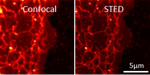
We have identified the causal connection between inhalation of nanoparticles, lipid wrapping and triggerring of the coagulation signal cascade in lung epithelium. The recently installed super-resolution STED microscope significantly contributed to this findings together with expertise transfer from UO and new proteomics techniques at UCD.
Download here
Published in PLoS ONE, 2018
Here we show that copper doped TiO2 nanotubes produce about five times more ·OH radicals as compared to undoped TiO2 nanotubes and that effective surface disinfection, determined by a modified ISO 22196:2011 test, can be achieved even at low intensity UVA light of 30 μW/cm2. The nanotubes can be deposited on a preformed surface at room temperature, resulting in a stable deposition resistant to multiple washings. Up to 103 microorganisms per cm2 can be inactivated in 24 hours, including resistant strains such as Methicillin-resistant Staphylococcus aureus (MRSA) and Extended-spectrum beta-lactamase Escherichia coli (E. coli ESBL).
Download here
Published in Viruses, 2018
To further refine the model of the molecular mobility on the HIV-1 surface, we herein investigated the relative importance of membrane composition and curvature in simplified model membrane systems, large unilamellar vesicles (LUVs) of different lipid compositions and sizes (0.1–1 µm), using super-resolution stimulated emission depletion (STED) microscopy-based fluorescence correlation spectroscopy (STED-FCS). Establishing an approach that is also applicable to measurements of molecule dynamics in virus-sized particles, we found, at least for the 0.1–1 µm sized vesicles, that the lipid composition and thus membrane rigidity, but not the curvature, play an important role in the decreased molecular mobility on the vesicles’ surface.
Download here
Published in European Biophysics Journal, 2019
We designed and tested several new orange and red fluorescent probes for live-cell STED nanoscopy. Three of the newly-synthesized probes exhibited improved resolution in STED nanoscopy with a 775 nm STED laser and two of these were also remarkably photostable, thus being perfectly suited for STED nanoscopy. The best overall results were assigned to the probe MePyr500, which was also not cytotoxic, making it an excellent candidate for live-cell STED nanoscopy.
Download here
Published in Journal of Biophonotics, 2020
Download here
Published in Applied Physics A, 2020
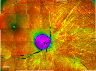
In our work, compact fiber-laser based on high-speed gain-switched laser diode has been shown to achieve adaptable/independently tunable repetition rate and energy per pulse allowing coupled fluorescence lifetime ophthalmic diagnostics via two-photon excitation and phototherapy via laser-induced photodisruption on a local molecular environment in a complex ex vivo retinal tissue.
Download here
Published in ACS Photonics, 2020
In this paper, we investigate how different STED confinement modes reduce the spurious background contributions from undepleted areas of the excitation in 3D FCS, thus increasing the signal quality and ultimately the resolution. By simulations as well as experiments with fluorescent probes in solution and in cells, we demonstrate that the coherent-hybrid (CH) depletion pattern created by a bivortex phase mask reduces background most efficiently and thus provides superior signal quality under comparable reduction of the observation volume.
Download here
Published in Advanced Materials, 2020
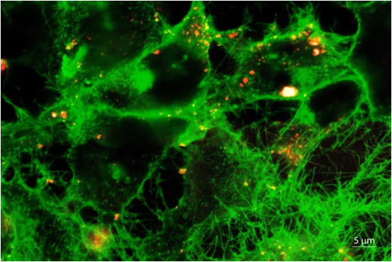
The prediction of diseases associated with nanomaterials is currently hampered by an incomplete understanding of the underlying mechanisms. Together with international collaborators, we discovered nanomaterial quarantining and counteracting nanomaterial cycling, which allowed us to incorporate these main modes of cellular response into a mechanistic model and predict in vivo inflammation solely on animal-free in vitro tests. The paper was published in the prestigious journal Advanced Materials and selected as the frontispiece of the journal issue.
Download here
Published in Materials, 2021
In our study, new insights of titanium alloy debris-tissue interaction were revealed by the implementation of label-free high-resolution correlative microscopy approaches: hybrid optical fluorescence and reflectance micro-spectroscopy, scanning electron microscopy (SEM), Energy-dispersive X-ray Spectroscopy (EDS), helium ion microscopy (HIM) and micro-particle-induced X-ray emission (micro-PIXE). Micron-sized wear debris were found as the main cause of the tissue oxidative stress exhibited through lipopigments accumulation in the nearby lysosome. This may explain the indications of chronic inflammation from prior histologic examination.
Download here
Published in FEBS Letters, 2021
To disentangle the elusive lipid–protein interactions in T-cell activation, we investigate how externally imposed variations in mobility of key membrane proteins (T-cell receptor [TCR], kinase Lck, and phosphatase CD45) affect the local lipid order and protein colocalisation. Using spectral imaging with polarity-sensitive membrane probes in model membranes and live Jurkat T cells, we find that partial immobilisation of proteins (including TCR) by aggregation or ligand binding changes their preference towards a more ordered lipid environment, which can recruit Lck. Our data suggest that the cellular membrane is poised to modulate the frequency of protein encounters upon alterations of their mobility, for example in ligand binding, which offers new mechanistic insight into the involvement of lipid-mediated interactions in membrane-hosted signalling events.
Download here
Published in Biomedical Optics Express, 2021
In our study, we present a proof-of-concept development of an algorithm to quantify laser-induced tissue changes on the basis of fluorescence lifetime and fluorescence intensity parameters. Using this algorithm, we can monitor and control the degree of laser-induced damage during theranostic procedures in real time.
Download here
Published in Nanotoxicology, 2021
The assessment of a nanoparticles’ mechanism of toxicity often depends on tracking the nanoparticles in cells or tissues using non-invasive advanced optical methods, such as live-cell fluorescence microscopy. This requires stable labeling of nanoparticles with fluorescent dyes - a process which, if not carefully controlled, can result in experimental artefacts that stem from the presence of unbound fluorophores and inadvertently altered chemical and physical properties of the nanomaterial.
Download here
Published in Nature Chemical Biology, 2023
The role of lipid membrane domains in the activation of immune cells remains elusive. New microscopy data on B cell signaling support a mechanism in which lipid membrane domains consolidate upon B cell receptor clustering.
Download here
Published in Nature methods, 2024
High laser powers in fluorescence microscopy should be used with care; besides the well-known photobleaching, the dye’s emission spectrum can unexpectedly shift towards lower wavelengths (“photoblueing”). The fluorescence signal is consequently detected in another spectral channel attributed to another dye, possibly leading to erroneous experimental conclusions. Detailed characterization of this phenomenon was done by our unique combination of a STED microscope equipped with a spectral detector.
Download here
Published in Bioorganic Chemistry, 2024
We synthesized and characterized a novel amphiphilic fluorescent label SHE-2N, which enables quick and long-lasting fluorescent labeling of the outer cell membranes of living cells. This new label for fluorescence microscopy is also suitable for high-resolution STED microscopy and two-photon microscopy due to its high photostability and appropriate spectral characteristics.
Download here
Published in Nanotoxicology, 2024
Currently, the limited models of sporadic Alzheimer’s disease fail to replicate all pathological hallmarks of the disease, making it difficult to understand the role of known environmental risk factors, such as exposure to air pollution, in the development of Alzheimer’s disease. Here, we report that exposure of the differentiated human neuroblastoma SH-SY5Y cell line to iron oxide and diesel exhaust particles (but not exposure to CeO2 nanoparticles) leads to formation of extracellular amyloid beta-containing plaques and decreased neurite length. This neurodegenerative phenotype is consistent with in vivo research and replicates all pathological hallmarks of Alzheimer’s disease, making the proposed in vitro model a useful tool for mechanistic investigations of environmental causes of Alzheimer’s disease.
Download here
Published in Nature Communications, 2024
Whole-genome duplications (WGDs) are a genomic event that enhances cancer metastasis and is associated with more aggressive cancers and shorter patient survival. We here show that cancers from self-reported Black patients exhibit a significantly higher frequency of WGD events compared to tumors from self-reported white patients. Moreover, their cancers also exhibit mutational signatures consistent with exposure to combustion byproducts. We demonstrate that these carcinogens are capable of inducing whole-genome duplications in cell culture.
Download here
Published:
We investigate basic molecular mechanisms of the blood coagulation cascade. In collaboration with Barry R. Lentz’s laboratory from University of North Carolina at Chapel Hill and with prof Rinku Majumders’ laboratory from the Louisiana State University School of Medicine in New Orleans, both recognized experts in the field of blood coagulation, Tilen Koklič showed heretofore yet unknown molecular mechanisms regarding regulation of factor Xa and of the prothrombinase (fXa-fVa) complex (Biochemical Journal, IF 4.40, Biophysical Journal, IF 4,32; Biochemistry, IF 3.56).
Published:

Published:

Published:

Published:

Published:

Published:
We are introducing adaptive acquisition schemes and computer vision algorithms to improve the efficiency of advanced optical techniques and reduce operators’ bias in the collected data. Within a national research project ARRS J7-2596, we collaborate with
Published:

Published:
Standardly equiped cell laboratory is placed in a clean room (ISO4/5) to ensure the most controllable environment to seed and grow cell lines. A clean chemical laboratory (ISO5) nearby ensures the most efficient hadnling during preparation and basic characterization of the samples. Finally, at the end of the air flow of the clean rooms, there is nano laboratory (ISO 6) for handling of nanomaterials and cleaning.
Published:
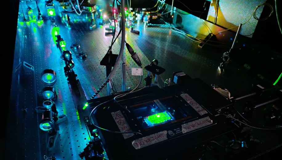
Optical setups are placed in the clean optical laboratory (ISO 6) together with modular platform for development of medical diagnostic technologies to ensure controllable and adaptable measurement environment. A small preparation lab enables final modification of the samples before optical measurements.
Published:
The home-made modular spinning-disk-based confocal fluroescence microscope with the ability of spectral detection for fast spectrally resolved imaging of environmentally sensitive probes, structures and interactions on the scales around 200 nm with spectral resolution of 1 nm. NIR optical tweezers enable contactless micro-manipulation.
Published:
The state-of-the-art modular 2-photon super-resolution 3D STED microscope, custom-built by Abberior Instruments, is equiped with several exciation and detection modalities for live-cell imaging of supramolecular structures on the scales down to 30 nm.
Univerza v Ljubljani / Mariboru ..., BSc/MSc.
Vedno smo veseli motiviranih študentov za izdelavo seminarjev, projektnih del in magistrskih nalog!
Univerza v Ljubljani, Fakulteta za farmacijo, Laboratorijska biomedicina, BSc.
Predmet se izvaja v letnem semestru 3. letnika programa Laboratorijska biomedicina (I. bolonjska stopnja) na Fakulteti za farmacijo Univerze v Ljubljani.